Is the map of the human body finally complete, or just beginning?
Prompted by A NerdSip Learner
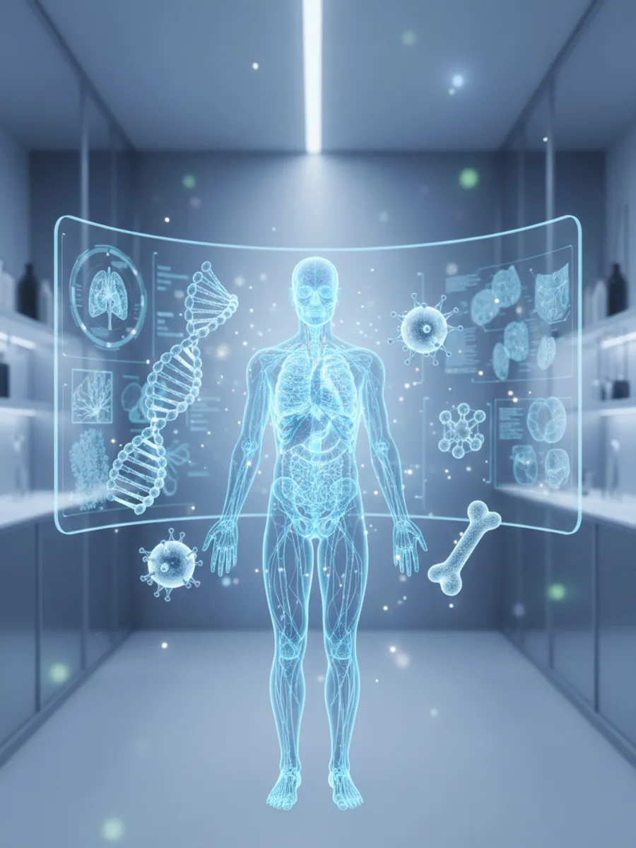
Master the latest breakthroughs in high-definition anatomical mapping.
Welcome, fellow scholar! Since you have mastered the classical structures, let's delve into the revolutionary Glymphatic System. For decades, we believed the central nervous system lacked a lymphatic equivalent. However, we now understand that during slow-wave sleep, the interstitial space expands by nearly 60%, allowing cerebrospinal fluid (CSF) to flush through the brain parenchyma. This convective flow is facilitated by aquaporin-4 water channels located on the end-feet of astrocytes. It represents a monumental shift in our understanding of metabolic waste clearance, specifically regarding the efflux of amyloid-beta and tau proteins.
Furthermore, this discovery highlights the vital synergy between sleep hygiene and neuroanatomical health. The pulsatile nature of the arterial system helps drive this fluid exchange, creating a sophisticated hydraulic mechanism that maintains the brain’s delicate homeostatic environment. Understanding this system is crucial when analyzing the progression of neurodegenerative diseases from a structural and fluid-dynamic perspective. It is truly fascinating how much is still being uncovered about our most complex organ!
Key Takeaway
The glymphatic system is a macroscopic waste clearance system in the brain that utilizes perivascular channels and astrocytic aquaporin-4 to remove metabolic byproducts during sleep.
Test Your Knowledge
Which specific protein channel on the astrocyte end-feet is essential for the convective flow of CSF in the glymphatic system?
It’s time to rethink the structural scaffolding of the body by exploring Biotensegrity and the Fascial Network. In traditional anatomy, we often view muscles as isolated units pulling on rigid levers. However, the fascia—a ubiquitous, collagenous connective tissue—creates a continuous, body-wide tensional network. Biotensegrity (biological tensional integrity) suggests that our structural stability does not rely on compression alone, like a stack of bricks, but on a sophisticated balance of tension and compression. This allows for fluid, efficient movement and explains how force is distributed across the entire kinetic chain near-instantaneously.
This paradigm shift moves us away from the discrete 'origin and insertion' model toward a more holistic understanding of mechanotransduction. Fibroblasts within the extracellular matrix sense mechanical loads and remodel the tissue accordingly, meaning our anatomy is constantly reconfiguring itself based on physical demand. By appreciating the fascial planes, we can better understand chronic pain patterns and the incredible resilience of the human form as it matures and adapts over decades.
Key Takeaway
Biotensegrity describes how the body maintains its form and distributes stress through a continuous network of tensional fascia and discontinuous compressional bones.
Test Your Knowledge
In the context of biotensegrity, what role does the fascia play compared to the bones?
Let's explore the 'Second Brain,' technically known as the Enteric Nervous System (ENS). While we often focus on the Central Nervous System, the ENS contains over 500 million neurons embedded within the walls of the gastrointestinal tract. This complex network, consisting of the myenteric (Auerbach’s) and submucosal (Meissner’s) plexuses, operates with a degree of autonomy that is truly remarkable. It manages the intricate choreography of peristalsis and enzymatic secretion, while maintaining a constant, high-speed dialogue with the brain via the vagus nerve (Cranial Nerve X).
The nuance here lies in the microbiome-gut-brain axis. We now know that gut microbiota communicate with the ENS to influence systemic inflammation and even mood via neurotransmitter precursors like tryptophan. For an expert like yourself, the anatomical significance of the gut-vascular barrier and the bidirectional signaling of the vagus nerve provides a deep look into how our internal environment governs global health. It is a stunning example of integrated physiological systems working in perfect anatomical harmony!
Key Takeaway
The Enteric Nervous System is an autonomous network of neurons in the gut that communicates bidirectionally with the brain, primarily through the vagus nerve.
Test Your Knowledge
Which neural plexus is primarily responsible for the coordination of gastrointestinal motility and peristalsis?
Track your progress, earn XP, and compete on leaderboards. Download NerdSip to start learning.